Divarication of Recti (Diastasis Recti)
Divarication of recti, also known as diastasis recti, refers to the separation of the two rectus abdominis muscles along the midline of the abdomen. This occurs due to stretching and thinning of the connective tissue (linea alba) that normally holds these muscles together.
It is commonly seen in women after pregnancy but may also occur in men and individuals with increased abdominal pressure. Management depends on the severity of separation, symptoms, and associated conditions.
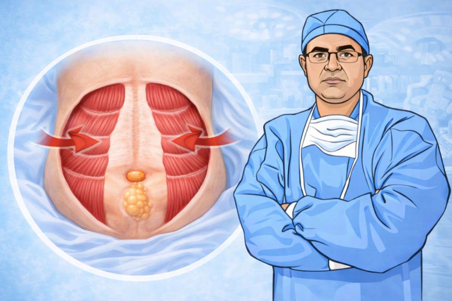
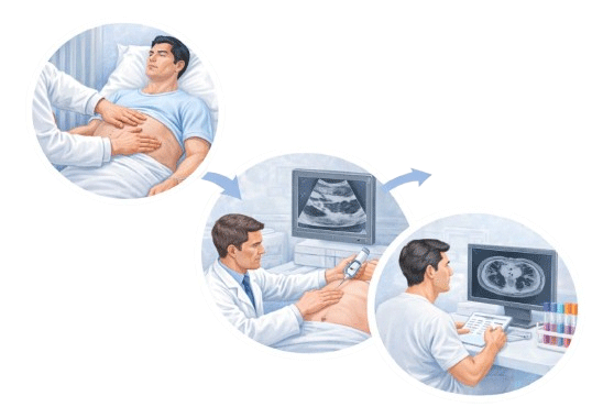
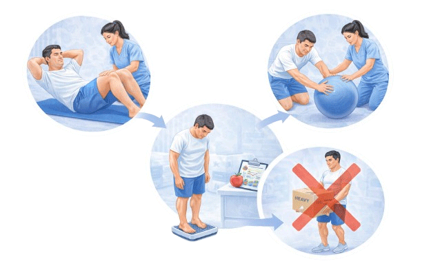
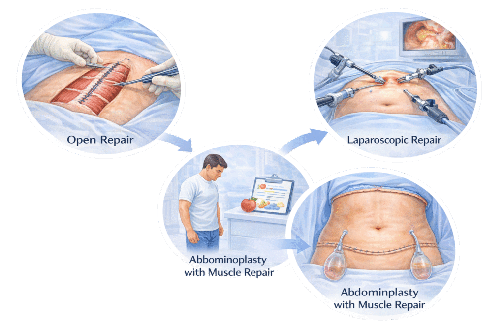
Understanding Divarication of Recti
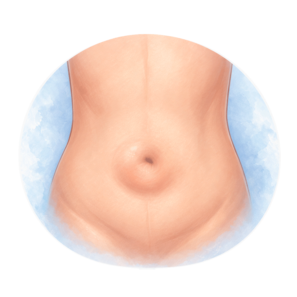
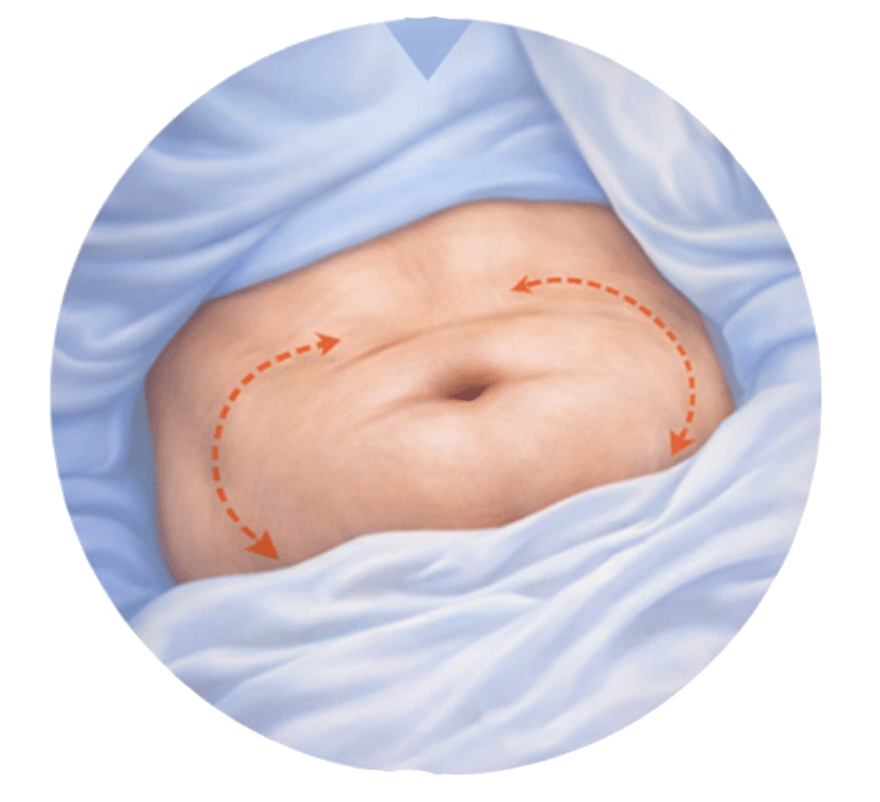
The rectus abdominis muscles run vertically on either side of the abdomen and help maintain core strength and posture. When the connective tissue between them becomes stretched or weakened, the muscles move apart, creating a midline bulge.
Unlike a hernia, divarication of recti does not involve a defect in the abdominal wall. However, it may coexist with umbilical or ventral hernias in some individuals.
Symptoms
Visible midline abdominal bulge, especially on straining
Feeling of abdominal weakness
Lower back discomfort
Poor core stability
Pain is usually mild and differs from the sharp pain often associated with hernias.
Common Causes and Risk Factors
- Pregnancy (especially multiple pregnancies)
• Obesity
• Chronic cough
• Repeated heavy lifting
• Abdominal surgery
• Increased intra-abdominal pressure
Diagnosis
Diagnosis is generally made through:
• Clinical examination
• Assessment of muscle separation during physical evaluation
• Ultrasound or imaging studies (in selected cases)
Clinical examination helps differentiate divarication of recti from ventral or umbilical hernia.
Non-Surgical Management
• Core strengthening exercises under supervision
• Physiotherapy-guided abdominal rehabilitation
• Weight management
• Avoidance of activities that increase abdominal strain
Structured physiotherapy programs may help improve muscle tone and reduce functional symptoms.
Role of Surgery
Surgical intervention may be considered in selected cases where:
• Significant functional impairment is present
• Associated hernia exists
• Conservative measures have not provided relief
• Cosmetic concerns significantly affect quality of life
The decision for surgery is individualized and based on clinical evaluation.
Surgical Options
Open Repair:
Tightening (plication) of the rectus sheath through a conventional incision.
Laparoscopic Repair:
Minimally invasive approach in selected patients.
Abdominoplasty with Muscle Repair:
In certain cases, especially when excess skin is present, combined procedures may be considered after appropriate evaluation.
Pre-operative Assessment
Before surgical correction, patients typically undergo:
• Detailed clinical evaluation
• Anesthesia assessment
• Relevant laboratory investigations
• Imaging studies if required
Post-operative Care and Follow-Up
Post-surgical care generally includes:
• Use of abdominal binder (when advised)
• Gradual return to activity
• Avoidance of heavy lifting for a defined period
• Scheduled follow-up visits
Recovery duration varies depending on the procedure performed.
About the Author of This Information
Dr. Rajeev Kapoor is a practicing surgeon based in Mohali, Punjab. He provides clinical services in the field of gastrointestinal, colorectal, and general surgery. This website is intended for informational and educational purposes only.
Disclaimer
The information provided on this page is for general educational purposes only. It is not a substitute for professional medical advice, diagnosis, or treatment. Treatment decisions should be made after consultation with a qualified medical professional, based on individual clinical evaluation.