blog

Early Sign of Colon Cancer
Cancer, blog

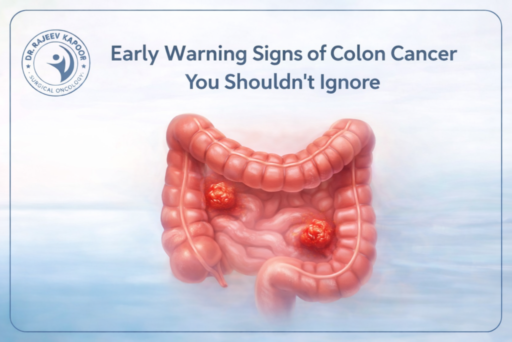
Early Warning Signs of Colon Cancer You Shouldn’t Ignore

Introduction Colon cancer, also known as colorectal cancer, is among the top five cancers affecting men and women in India. Yet, most patients are diagnosed only when the disease has already advanced. According to Dr. Rajeev Kapoor, a renowned gastrointestinal and cancer surgeon in Chandigarh, “Early detection is the key to saving lives. Awareness of the initial warning signs can make all the difference.” 1️⃣ Change in Bowel Habits One of the earliest indicators of colon cancer is a persistent change in bowel movements—such as diarrhea, constipation, or a feeling that the bowel does not empty completely. If such symptoms last more than two weeks, they should not be ignored. 💡 Tip from Dr. Kapoor: “If your bowel habits have changed and do not respond to routine diet adjustments, consult your doctor for evaluation.” 2️⃣ Blood in Stool or Rectal Bleeding The appearance of bright red or dark blood in stool may signal bleeding from the lower intestine. Though hemorrhoids are a common cause, hidden bleeding could also indicate colorectal cancer. Periodic screening can detect such bleeding early, even before visible symptoms appear. 3️⃣ Unexplained Weight Loss and Fatigue Unintentional weight loss, fatigue, or persistent weakness could point to a deeper issue. Cancer cells consume more energy, while slow internal bleeding can lead to iron-deficiency anemia, which often manifests as chronic tiredness. 4️⃣ Abdominal Pain and Bloating Patients may experience cramping, gas, or discomfort in the lower abdomen. These symptoms can be mistaken for IBS (Irritable Bowel Syndrome) but, when persistent, require a colonoscopy to rule out malignancy. 5️⃣ Family History and Genetic Risk Individuals with a family history of colon or rectal cancer—or conditions such as Lynch syndrome or familial adenomatous polyposis (FAP)—have a significantly higher risk. Dr. Kapoor recommends screening colonoscopy starting at age 45 or even earlier for those with genetic predispositions. Why Early Detection Matters When colon cancer is caught early, the survival rate can exceed 90 %. Screening methods like colonoscopy, stool occult blood tests, and CT colonography can identify precancerous polyps that can be removed before they turn cancerous. 🩺 “Every colon cancer begins as a small, silent polyp. Detecting it before it turns malignant can save a life,” emphasizes Dr. Kapoor. Prevention and Lifestyle Tips Eat a fiber-rich diet (fruits, vegetables, whole grains) Limit red and processed meat Maintain a healthy weight and regular exercise routine Avoid smoking and excess alcohol Schedule periodic screenings after 45 (or earlier if at risk) Conclusion Colon cancer is preventable, treatable, and—even in many cases—curable when detected early. Awareness of these warning signs and timely medical evaluation can dramatically improve outcomes. If you or a loved one experience any of these symptoms, don’t delay. 

soft tissue sarcoma
Sarcoma, blog, Cancer

Soft Tissue Sarcoma: Understanding This Rare but Treatable Cancer

Soft tissue sarcoma (STS) is a rare group of cancers that arise from the body’s connective tissues—muscles, fat, nerves, and blood vessels. Though uncommon, these tumors can occur anywhere in the body and often remain silent until they reach an advanced stage. “Sarcomas are challenging because they can grow quietly for months. But with today’s imaging and surgical advances, early intervention leads to excellent results,” explains Dr. Rajeev Kapoor, senior cancer and gastrointestinal surgeon based in Chandigarh. 1️⃣ What Is Soft Tissue Sarcoma? Soft tissue sarcomas comprise over 70 different subtypes, including liposarcoma, leiomyosarcoma, synovial sarcoma, and angiosarcoma. They account for only about 1 % of adult cancers yet can occur at any age. These tumors develop from cells that form structural tissues of the body — making them capable of appearing in the limbs, abdomen, chest, or head-and-neck region. 2️⃣ Key Symptoms You Should Watch For A painless, enlarging lump anywhere in the body Deep-seated swelling or mass > 5 cm that persists Unexplained pain or pressure when the tumor presses against nerves Reduced mobility if located near joints or muscles Unexplained weight loss or fatigue in later stages “Any lump that grows steadily or returns after removal must never be ignored,” warns Dr. Kapoor. 3️⃣ Diagnosis and Imaging Techniques A precise diagnosis requires a multidisciplinary approach: MRI Scan: Determines tumor size and relation to surrounding structures CT Scan: Assesses spread to lungs or other organs Core-Needle Biopsy: Essential for histopathological confirmation PET-CT: Evaluates metastatic disease and treatment planning Dr. Kapoor stresses the importance of getting biopsies performed only by trained oncological surgeons, as improper procedures can complicate later surgeries. 4️⃣ Treatment Options Modern treatment for soft tissue sarcoma is both effective and customized: a. Surgery – Cornerstone of Treatment Wide local excision removing tumor with clear margins Limb-sparing techniques preserve function without amputation b. Radiation Therapy Given before or after surgery to reduce recurrence risk Image-guided and intensity-modulated radiation (IMRT) minimizes damage to normal tissue c. Chemotherapy and Targeted Therapy Drugs like Doxorubicin and Ifosfamide for aggressive subtypes Targeted molecules and immunotherapy agents are showing remarkable promise in advanced cases “Our goal is to combine oncologic safety with functional preservation. With modern techniques, we can save limbs and lives,” adds Dr. Kapoor. 5️⃣ Survival and Prognosis When detected early and managed by a specialized team, soft tissue sarcoma can be successfully treated. The overall 5-year survival rate ranges between 65–80 %, depending on the tumor stage and type. Regular follow-up is crucial to monitor for recurrence or metastasis. 6️⃣ Early Detection and Awareness in India In India, sarcomas are often diagnosed late due to lack of awareness and misdiagnosis as benign lumps. Public education and early screening efforts — especially through campaigns like Sarcoma Awareness Month (July) — can significantly improve outcomes. Conclusion Soft tissue sarcoma may be rare, but it is not untreatable. Advances in diagnostics, precision surgery, and targeted therapies have dramatically improved prognosis. If you notice a persistent or unusual lump, consult a specialist without delay. Dr. Rajeev Kapoor, a trusted cancer and gastrointestinal surgeon in Chandigarh, leads a multidisciplinary team offering comprehensive care for sarcoma and other complex cancers.

diet to reduce your risk of colon cancer
blog, Cancer

Can Diet and Lifestyle Reduce Your Risk of Colon Cancer?

Colon cancer is increasingly affecting younger adults in India, largely due to modern dietary habits and sedentary lifestyles. But the good news is — many cases can be prevented through simple lifestyle changes. “Nearly 40% of colon cancers are linked to modifiable risk factors such as diet, obesity, and inactivity,” says Dr. Rajeev Kapoor, one of Chandigarh’s leading gastrointestinal and cancer surgeons. “Your plate and your lifestyle can be your first line of defense.” 1️⃣ The Link Between Diet and Colon Cancer Our colon is directly influenced by what we eat. Diets low in fiber and high in fat, processed meats, and refined carbohydrates can trigger inflammation and alter gut bacteria, paving the way for cancerous changes. High-risk foods include: Red meat (beef, pork, lamb) — increases exposure to carcinogenic compounds Processed meats (sausages, ham, bacon) — rich in nitrates and preservatives Refined sugars and junk food — promote obesity and insulin resistance On the other hand, diets rich in fiber, antioxidants, and omega-3 fatty acids can protect colon health. 2️⃣ Protective Foods That Help Prevent Colon Cancer Food Group Protective Role Examples Fiber-rich foods Speeds up bowel movement, reduces toxin exposure Whole grains, lentils, oats Cruciferous vegetables Contain sulforaphane, a cancer-fighting compound Broccoli, cabbage, cauliflower Leafy greens Rich in folate and antioxidants Spinach, kale, methi Probiotic foods Balance gut microbiota Curd, yogurt, kefir Omega-3 sources Reduce inflammation Fish, flaxseed, walnuts Fruits & citrus Vitamin C and flavonoids protect colon lining Oranges, papaya, apples “I always tell my patients — a colorful plate is a healthy plate. Each color represents a different protective nutrient,” notes Dr. Kapoor. 3️⃣ The Role of Lifestyle in Colon Cancer Prevention a. Regular Physical Activity At least 150 minutes of moderate exercise per week (like brisk walking, yoga, cycling) Physical activity helps maintain a healthy weight and improves bowel motility b. Weight Management Obesity, especially central abdominal fat, increases the risk of colon cancer by altering insulin and hormone balance. Maintaining a BMI below 25 significantly lowers risk. c. Quit Smoking and Limit Alcohol Both smoking and alcohol increase oxidative stress in colon cells, contributing to DNA damage. Moderate or avoid completely. d. Regular Screening and Checkups Screening is crucial even for those without symptoms, especially after age 45 or earlier if there’s a family history. Recommended tests include: Fecal occult blood test (FOBT) Colonoscopy (gold standard for early detection) CT colonography “Many people wait for symptoms before they act. Screening saves lives — it detects precancerous polyps before they become cancer,” emphasizes Dr. Kapoor. 4️⃣ The Indian Context: Changing Food Habits, Rising Risks The shift from traditional Indian diets to Westernized fast-food patterns has led to increased incidence of lifestyle diseases — including colon cancer. Urbanization, stress, and late-night eating further worsen digestive health. Promoting awareness about balanced diets, home-cooked meals, and mindful eating can be powerful public-health strategies in India. 5️⃣ Your Colon Health Checklist ✅ Eat at least 25–30 grams of fiber daily ✅ Include 2–3 servings of fruits & vegetables in every meal ✅ Avoid processed meats and limit red meat ✅ Exercise 30 minutes a day ✅ Schedule a screening colonoscopy after 45 ✅ Stay hydrated and manage stress Conclusion Colon cancer is not inevitable — it’s preventable. By making conscious food and lifestyle choices, you can dramatically reduce your risk and protect your digestive health for years to come. Dr. Rajeev Kapoor, a trusted gastrointestinal and cancer surgeon in Chandigarh, advocates for preventive healthcare through lifestyle awareness, early diagnosis, and modern surgical care. “Prevention is far more powerful than treatment — and it begins in your kitchen.”

Advances in Minimally Invasive Cancer Surgery
Cancer, blog

Advances in Minimally Invasive Cancer Surgery: How Technology Is Changing Outcomes

Cancer treatment has evolved dramatically in the last decade. The scalpel is no longer the only symbol of surgery — today, precision, technology, and faster recovery define success. “Our goal is not just to remove the tumor, but to restore quality of life as quickly as possible. That’s where minimally invasive surgery is transformative,” explains Dr. Rajeev Kapoor, senior gastrointestinal and cancer surgeon at Chandigarh. 1️⃣ What Is Minimally Invasive Cancer Surgery? Minimally invasive cancer surgery (MICS) refers to procedures performed through tiny incisions using cameras and specialised instruments, instead of large open cuts. Techniques include: Laparoscopic surgery Robotic-assisted surgery Thoracoscopic and endoscopic resections These approaches allow surgeons to access complex areas like the colon, stomach, or pancreas with remarkable precision — while minimizing trauma to healthy tissues. 2️⃣ Key Advantages for Patients Benefit Impact Smaller incisions Less pain and scarring Minimal blood loss Reduced transfusion risk Faster recovery Shorter hospital stay Lower infection rate Enhanced postoperative safety Early return to routine Better patient experience “In colon and gastrointestinal cancers, laparoscopic and robotic surgery can reduce hospital stay from 8–10 days to just 3–5 days,” shares Dr. Kapoor. 3️⃣ Robotic Surgery: The New Frontier Robotic-assisted systems such as Da Vinci Surgical Robot bring magnified 3D visualization and ultra-precise hand movements, translating the surgeon’s motions into micro-movements inside the body. This allows for: Millimeter-level precision in delicate dissections Reduced nerve damage in pelvic and rectal surgeries Improved oncological margins (complete tumor removal) Dr. Kapoor’s team employs these technologies in colorectal, hepatobiliary, and sarcoma resections, ensuring both oncological safety and functional preservation. 4️⃣ Applications in Gastrointestinal and Soft Tissue Cancers Colon & Rectal Cancer: Laparoscopic colectomy and robotic rectal surgery Stomach Cancer: Minimally invasive gastrectomy for early and advanced cases Liver and Pancreas: Keyhole resections with enhanced recovery programs Sarcoma: Limb-sparing resections using precision navigation tools “For many cancers, patients can walk within 24 hours and resume normal diet in two days. That’s the power of technology-led care,” notes Dr. Kapoor. 5️⃣ Safety and Oncological Outcomes Extensive global studies confirm that minimally invasive techniques achieve equal or better cancer-control outcomes compared to open surgery — provided they are performed by trained specialists. Postoperative recovery, immune response, and quality of life are significantly superior. 6️⃣ The Future of Cancer Surgery in India India is rapidly adopting robotics, fluorescence imaging, and AI-driven navigation tools. Dr. Kapoor foresees a future where precision oncology, powered by technology and data, makes cancer surgery safer, smarter, and more personalized. “We are entering an era where technology complements judgment — allowing us to heal, not just operate.” Conclusion Minimally invasive cancer surgery represents a revolution in cancer care — reducing pain, enhancing precision, and restoring dignity in recovery. For patients seeking advanced, evidence-based, and compassionate treatment, Dr. Rajeev Kapoor and his team in Chandigarh offer state-of-the-art surgical expertise backed by global standards.

Understanding Hernia
blog, Hernia

Understanding Hernia: Causes, Symptoms and When to Seek Surgery

A hernia often begins as a small, painless swelling but may gradually progress if not medically evaluated. In clinical terms, a hernia occurs when an internal organ or tissue protrudes through a weakened area of the muscle or connective tissue, leading to a visible or palpable bulge. According to Dr. Rajeev Kapoor, senior gastrointestinal and hernia surgeon based in Chandigarh, most hernias do not resolve on their own and require appropriate medical assessment to prevent complications. 1. What Exactly is a Hernia? Our abdominal wall is made of layers of muscles and connective tissues that hold organs in place. When this wall weakens due to age, strain, or surgery, internal tissue can protrude through — forming a hernia. Common Types of Hernia Type Location Common in Inguinal Hernia Groin area Men (most common type) Umbilical Hernia Around the navel Infants, obese adults Incisional Hernia Through a previous surgical scar Post-operative patients Hiatal Hernia Upper stomach, near diaphragm Adults over 40 yrs Femoral Hernia Upper thigh/groin crease Women (less common) 2. Why Do Hernias Occur? Several lifestyle and medical factors contribute to hernia formation: Chronic coughing or sneezing (COPD, allergies) Heavy lifting without core support Long-term constipation and straining during bowel movements Obesity or sudden weight gain Previous abdominal surgery or trauma Pregnancy (increases abdominal pressure) As Dr. Kapoor explains, hernias typically develop over time due to gradual weakening of tissues rather than appearing suddenly. 3. Symptoms That Require Attention Some hernias may remain asymptomatic in the early stages, while others may cause noticeable discomfort. Common symptoms include: A visible or palpable bulge that becomes more prominent on standing or coughing Mild to moderate pain, discomfort, or a burning sensation Pain during physical activity, bending, or lifting A feeling of heaviness or pressure in the affected area Warning Signs Requiring Medical Care Sudden severe pain at the hernia site Inability to push the bulge back in Nausea, vomiting, or abdominal distension Redness, hardness, or tenderness over the swelling These symptoms may indicate complications such as incarceration or strangulation of the hernia, which require immediate medical intervention. 5. Treatment Options for Hernia a. Conservative (Non-Surgical) Management In select cases where the hernia is small and asymptomatic, careful observation may be advised. However, it is important to note that the structural defect does not heal spontaneously. b. Surgical Management Surgery remains the definitive treatment for hernia repair. The commonly used approaches include: Open Hernia Repair: The weakened area is repaired through a surgical incision, often reinforced with mesh. Laparoscopic Hernia Repair: A minimally invasive approach using small incisions and camera-guided instruments. The choice of surgical technique depends on multiple factors such as hernia type, patient health status, and surgeon assessment. 6. Measures That May Reduce Risk While hernias cannot always be prevented, certain measures may help reduce risk: Maintaining a healthy body weight Using proper techniques during lifting Managing chronic cough or constipation Ensuring adequate nutrition, including sufficient protein intake Avoiding smoking, as it affects tissue healing Conclusion A hernia is a common medical condition that requires proper clinical evaluation. Although it may appear minor initially, delayed assessment can lead to complications. Early diagnosis and appropriate management help ensure safer outcomes and recovery. This information is intended to support patient awareness and does not replace professional medical consultation. Frequently Asked Questions (FAQs): Understanding Hernia 1. What is a hernia? A hernia occurs when an internal organ or tissue pushes through a weak area in the muscle or connective tissue that normally holds it in place. This often appears as a visible or palpable bulge. 2. Are hernias common? Yes. Hernias are a common medical condition and can affect people of all ages, including infants and older adults. Some types are more frequent in men, while others are more common in women. 3. What causes a hernia? Hernias usually develop due to a combination of muscle weakness and increased pressure inside the abdomen. Contributing factors may include: Age-related muscle weakness Heavy lifting or physical strain Chronic coughing or constipation Obesity Pregnancy Previous abdominal surgery 4. Can a hernia heal on its own? No. The muscle defect causing a hernia does not close on its own. While symptoms may remain mild for some time, the hernia itself persists unless treated surgically. 5. What are the common symptoms of a hernia? Symptoms may include: A bulge that becomes more noticeable on standing or coughing Discomfort, pain, or a burning sensation Heaviness or pressure in the affected area Some hernias may cause little or no discomfort initially. 6. Are all hernias painful? Not necessarily. Many hernias are painless in the early stages. Pain may develop as the hernia enlarges or if complications arise. 7. When is a hernia considered an emergency? A hernia requires urgent medical attention if there is: Sudden severe pain A hard or tender bulge that cannot be pushed back Nausea, vomiting, or abdominal swelling These symptoms may indicate incarceration or strangulation of the hernia. 8. What are the different types of hernia? Common types include: Inguinal hernia (groin area) Umbilical hernia (around the navel) Incisional hernia (at a previous surgical site) Hiatal hernia (upper stomach near the diaphragm) Femoral hernia (upper thigh or groin crease) 9. How is a hernia diagnosed? Diagnosis is usually based on medical history and physical examination. Imaging tests such as ultrasound, CT scan, or endoscopy may be advised in certain cases. 10. Do all hernias require surgery? Not all hernias require immediate surgery. In some small or asymptomatic cases, observation may be recommended. However, surgery is the only definitive treatment to repair the muscle defect. 11. What are the treatment options for hernia? Treatment options include: Observation in selected cases Surgical repair, either through open surgery or laparoscopic (minimally invasive) techniques The choice depends on hernia type, symptoms, and patient health. 12. Is laparoscopic surgery better than open surgery? Both methods are effective. Laparoscopic surgery may offer faster recovery and smaller scars for suitable patients, while open surgery may be preferred in certain situations. The decision is

Fournier’s Gangrene Treatment in Chandigarh
blog

Fournier’s Gangrene: A Life-Threatening Soft Tissue Infection Requiring Urgent Surgical Care

Fournier’s gangrene is a rare but extremely serious infection affecting the genitals, scrotum, and perineum. It is a form of necrotising fasciitis, where bacteria destroy tissues rapidly beneath the skin. Patients can deteriorate within hours due to septicemia (blood poisoning), and delayed treatment increases the risk of permanent disability and even death. With mortality rates ranging from 20–40% in early presenters and up to 50% or more in late or critically ill patients, this condition is one of the most urgent emergencies in general surgery. Early recognition and immediate surgical treatment save lives. What Exactly Is Fournier’s Gangrene? Fournier’s gangrene begins when bacteria enter through a small wound, boil, abscess, perianal infection, or urinary tract infection. These bacteria spread quickly in the deeper tissues, releasing toxins that cause: tissue death (necrosis) gas formation under the skin rapid extension into surrounding areas The dangerous part is that the surface may look mild while the deeper tissues are already severely damaged. This silent progression is why Fournier’s gangrene must be treated as a surgical emergency. Who Is at Risk? Anyone can develop this infection, but certain conditions significantly increase the risk: • Diabetes (poorly controlled or undiagnosed) • Obesity • Smoking or heavy alcohol use • Kidney or liver disease • Immunosuppression (cancer therapy, steroids, HIV) • Recent surgery or trauma in the genital/perineal area • Chronic skin infections or boils Many patients discover they have uncontrolled diabetes for the first time after presenting with this condition. Symptoms You Should Never Ignore Fournier’s gangrene often starts subtly but worsens fast. Seek emergency medical care if you notice: Sudden or severe pain in the scrotum, penis, or perineum Rapid swelling, redness or spreading warmth Fever, chills, extreme fatigue Dark patches of skin — purple, grey, or black Foul-smelling discharge Crackling sensation under the skin (due to gas-producing bacteria) Pain that feels far worse than what the skin looks like These red flags may indicate the infection is spreading deep into the tissues. Diagnosis: How Doctors Confirm the Condition A diagnosis is made using: • Clinical examination • Blood tests (showing infection, kidney stress, metabolic changes) • Ultrasound or CT scan to check spread and gas formation • Immediate surgical evaluation Because time lost is life lost, treatment begins immediately, without waiting for complete reports. Resuscitation is done in an ICU according to patients general condition. Treatment: Fast, Aggressive, and Life-Saving Managing Fournier’s gangrene requires a coordinated, aggressive approach including emergency surgery, antibiotics, and critical care support. 1. Emergency Surgical Debridement This is the most important part of treatment. Surgeons remove all dead, infected, and dying tissue. Surgery is performed as soon as the diagnosis is suspected. Because the infection spreads rapidly, most patients require multiple debridements over several days. Early repeated surgery prevents further spread and reduces mortality. Although extensive, these procedures are absolutely lifesaving. 2. Broad-Spectrum Intravenous Antibiotics Strong IV antibiotics are given immediately to target all likely bacteria — aerobic and anaerobic. Treatment continues for days or weeks depending on progress. 3. ICU Care and Stabilisation Many patients arrive in septicemia, a life-threatening state where bacteria enter the bloodstream. ICU management may include: • stabilising blood pressure • supporting kidney and lung function • managing fluids and electrolytes • oxygen support • insulin infusions for blood sugar control • treatment for septic shock if present Early ICU involvement significantly improves outcomes. 4. VAC Dressing (Negative Pressure Wound Therapy) After debridement, a VAC dressing (vacuum-assisted closure) helps: • remove infected fluid • reduce bacterial load • speed up healing • improve tissue granulation • reduce dressing frequency • prepare the wound for reconstruction VAC therapy is now a standard part of advanced wound management for Fournier’s gangrene. Reconstruction After the Infection Is Controlled Once the infection stops and the wound is clean, reconstructive surgery begins. Reconstruction is tailored to the tissue lost and may involve: • Split-thickness skin grafts • Local flaps • Scrotal reconstruction • Penile skin reconstruction • Perineal defect closure Most patients regain excellent function and appearance after reconstruction. Recovery and Long-Term Outlook With early and aggressive treatment: • Most patients recover well • Wounds heal over several weeks to months • Follow-up ensures diabetes control and wound healing Recovery time depends on: • how early treatment began • the amount of tissue removed • severity of septicemia • underlying health conditions Patients who present late may require longer reconstructive and rehabilitative support. When to Seek Emergency Care Visit the nearest emergency department immediately if you notice: • sudden severe genital or perineal pain • black or discoloured skin patches • rapid swelling or redness • fever or chills with genital symptoms • foul-smelling discharge • a rapidly worsening boil or abscess Do not wait. Fournier’s gangrene is a medical emergency. Can Fournier’s Gangrene Be Prevented? You can reduce your risk by: • maintaining good diabetes control • treating boils or urinary infections early • avoiding smoking and excessive alcohol • keeping the genital area clean and dry • managing chronic illness responsibly • seeking medical care for any non-healing wound in the groin Prevention focuses on recognising problems early and treating them promptly.

Colon Cancer Patient Story
blog, Cancer

Real Colon Cancer Patient Story: Early Detection to Complete Recovery

Mr. Khanna’s story began with two signs that should never be ignored—persistent abdominal pain and blood in the stool. These symptoms are common in many conditions, but they are also early warning signs of colorectal cancer. His doctor advised immediate tests, including a CT scan and then a colonoscopy. The colonoscopy showed a growth in the right side of the colon that looked cancerous. He met me soon after. I explained that colon cancer often starts as a small polyp that slowly grows over years. Many people have no symptoms until the tumour becomes large. In Mr. Khanna’s case, the safest and most effective treatment was surgery. He underwent a laparoscopic right hemicolectomy, which is the standard operation for cancers on the right side of the colon. In this procedure, the part of the bowel containing the tumour is removed, along with the blood vessels and lymph nodes that drain that area. This is important because cancer cells can travel through these channels. We removed around 30 lymph nodes, which gave us a clear picture of how far the disease had spread. Laparoscopic surgery, done through small cuts, usually means less pain, quicker recovery, and faster return to normal activity. Mr. Khanna recovered well and went home in five days. Thirteen days later, the final pathology report came. The cancer had been completely removed with clear margins, meaning no cancer cells were left behind at the edges. All his lymph nodes were negative for cancer. This placed him in Stage II colon cancer. However, the tumour had reached the outer layer of the bowel wall, known as the serosa. When a tumour grows this deep, there is a higher chance—though still small—of cancer cells escaping into the bloodstream. Because of this “high-risk” feature, chemotherapy was advised to lower the risk of the cancer returning. He had a chemo port inserted in his chest. Chemotherapy for colon cancer usually involves drugs like oxaliplatin and capecitabine/5-FU, given over three months in Stage II high-risk cases. These medicines target any microscopic cancer cells that surgery cannot see. The first few cycles went as expected—tiredness, nausea, changes in taste. After the third cycle, he developed fever and a rash. Tests showed herpes zoster (shingles), an infection that can occur when immunity is low during chemotherapy. He was treated with antiviral medication, recovered fully, and safely completed the rest of his chemotherapy. Today, Mr. Khanna has finished treatment and is doing well. For colon cancer survivors, follow-up care is very important. His plan includes: • Blood tests, including CEA (a tumour marker) • Periodic CT scans to check the abdomen and chest • Regular colonoscopies to detect new polyps early • Lifestyle advice—healthy diet, regular exercise, weight control, and avoiding smoking His story shows how colon cancer is diagnosed and treated today. Early symptoms may be mild. Early surgery, when possible, offers the best chance of cure. Chemotherapy adds extra protection when the tumour shows high-risk features. By sharing his journey, we hope people understand the importance of recognising symptoms, seeking medical help early, and trusting modern treatment pathways. With timely care, most patients with early-stage colon cancer can expect excellent long-term outcomes and a full return to normal life.

Laparoscopic Hernia Repair
blog, Hernia

Laparoscopic Hernia Repair: A Faster, Safer, and Scar-Free Option

Advances in surgical techniques have significantly changed the management of hernias. One such development is laparoscopic hernia repair, a minimally invasive approach that allows repair through small incisions and is associated with faster recovery compared to traditional open surgery. According to Dr. Rajeev Kapoor, senior gastrointestinal and laparoscopic surgeon in Chandigarh based in Chandigarh, laparoscopic techniques have improved patient comfort and reduced hospital stay in appropriately selected cases.   1️⃣ What Is Laparoscopic Hernia Repair? Laparoscopic hernia repair, also known as keyhole surgery, involves repairing the hernia through three tiny incisions (each about 0.5–1 cm). A thin tube with a camera (laparoscope) is inserted to provide a magnified, high-definition view of the internal structures. Using specialized instruments, the surgeon gently pushes the protruding tissue back into place and reinforces the weak spot with a surgical mesh, ensuring long-term strength. 2️⃣ How It Differs from Open Surgery Aspect Open Hernia Repair Laparoscopic Hernia Repair Incision Size 6–10 cm (large cut) 0.5–1 cm (keyhole) Pain & Recovery Moderate to high pain, longer rest Minimal pain, rapid recovery Hospital Stay 2–4 days Same-day discharge (24 hrs max) Scarring Visible scar Nearly invisible Recurrence Rate Slightly higher Very low when done by experts Return to Work 10–15 days 3–5 days The choice between open and laparoscopic surgery depends on the type of hernia, patient health, and clinical evaluation. 3️⃣ Who Can Benefit from Laparoscopic Hernia Surgery? Laparoscopic repair is suitable for: Inguinal (groin) hernias — the most common type in men Bilateral hernias (both sides) Recurrent hernias (after open surgery) Umbilical and incisional hernias Obese patients, where open surgery carries more risk However, not all hernias are suitable for a laparoscopic approach. Large, complicated, or strangulated hernias require careful assessment before deciding on the surgical method. 4️⃣ How the Procedure Is Performed General anesthesia is given for patient comfort. Small incisions are made near the navel. A laparoscope provides a magnified view of the hernia and surrounding tissue. The protruding tissue is gently repositioned inside the abdomen. A medical-grade mesh is placed over the weak area to reinforce the wall. The incisions are closed with absorbable sutures — leaving minimal or no visible scar. The procedure duration varies depending on the type and complexity of the hernia. 5️⃣ Recovery and Post-Surgery Care Walking: Within 4–6 hours post-surgery Diet: Light meals on the same day Return to Work: Usually within 3–5 days Exercise/Lifting: Avoid heavy weights for 4–6 weeks Follow-Up: Regular reviews ensure healing and prevent recurrence Recovery timelines may differ based on individual health and the nature of the surgery. 6️⃣ Potential Benefits of Laparoscopic Hernia Repair When clinically appropriate, laparoscopic repair may offer: Smaller incisions and reduced tissue trauma Shorter hospital stay Faster return to daily activities Lower risk of wound-related complications Improved cosmetic outcome All surgical procedures carry some degree of risk, which should be discussed in detail with the treating surgeon. 7️⃣ Role of the Treating Surgeon Successful hernia management depends on accurate diagnosis, appropriate surgical planning, and post-operative care. Surgeons trained in both open and laparoscopic techniques can determine the most suitable approach based on patient-specific factors. Dr. Rajeev Kapoor is a senior gastrointestinal and laparoscopic surgeon who manages hernia cases as part of his clinical practice, with emphasis on patient safety and evidence-based decision-making. Conclusion Laparoscopic hernia repair is a well-established surgical option that may be suitable for many patients, offering the advantages of minimally invasive surgery when appropriately indicated. Early medical evaluation and informed discussion with a qualified surgeon are essential for optimal outcomes. This article is intended for general patient education and should not replace personalised medical consultation. Frequently Asked Questions (FAQs): Laparoscopic Hernia Repair 1. What is laparoscopic hernia repair? Laparoscopic hernia repair is a minimally invasive surgical technique in which a hernia is repaired using small incisions, a camera (laparoscope), and specialised instruments. The weakened area of the abdominal wall is reinforced, often using a surgical mesh. 2. How is laparoscopic hernia surgery different from open surgery? In laparoscopic surgery, multiple small incisions are used instead of a single larger incision. This approach generally results in less tissue trauma, smaller scars, and faster recovery in suitable patients. The choice of method depends on clinical evaluation. 3. Is laparoscopic hernia repair safe? Laparoscopic hernia repair is a well-established and commonly performed procedure. Like all surgeries, it carries some risks, which should be discussed with the surgeon before the procedure. 4. Who is a suitable candidate for laparoscopic hernia repair? Laparoscopic repair may be considered for patients with: Inguinal hernias Bilateral hernias Recurrent hernias Umbilical or incisional hernias Suitability depends on the size of the hernia, patient health and surgeon assessment. 5. Can all hernias be treated laparoscopically? No. Very large, complicated, or strangulated hernias may require open surgery. The decision is made after careful clinical evaluation and imaging, if required. 6. Does laparoscopic hernia surgery require general anaesthesia? Yes. Laparoscopic hernia repair is typically performed under general anaesthesia to ensure patient comfort and safe surgical access. 7. How long does laparoscopic hernia surgery take? The duration varies depending on the type and complexity of the hernia. Many procedures are completed within one to two hours. 8. Will a mesh be used during laparoscopic hernia repair? In most cases, a medical-grade surgical mesh is used to reinforce the weakened area. Mesh use reduces the risk of recurrence and provides long-term strength to the repair. 9. Is mesh placement safe? Surgical mesh is widely used in hernia repair and has been extensively studied. As with any implant, mesh-related risks exist and should be discussed with the surgeon prior to surgery. 10. How long is the hospital stay after laparoscopic hernia surgery? Many patients are discharged on the same day or within 24 hours, depending on recovery, pain control, and overall health. 11. What is the recovery time after laparoscopic hernia repair? Most patients resume light activities within a few days. Full recovery, including internal healing, may take several weeks. Recovery timelines vary individually. 12. Will there be visible

Recurrent Biliary Colic
blog

Recurrent Biliary Colic in a Diabetic Patient: Why Early Surgery Matters

Gallstones are common, but when they begin causing repeated episodes of abdominal pain, timely treatment becomes crucial—especially in patients with diabetes. Here is a real case shared by Dr. Rajeev Kapoor, highlighting the importance of early intervention in a 40-year-old woman with type 2 diabetes and recurrent biliary colic. What Is Biliary Colic? Biliary colic is the sudden, sharp pain caused when a gallstone temporarily blocks the cystic duct—the channel that connects the gallbladder to the bile duct. The pain usually: Comes on suddenly Occurs after oily or heavy meals Lasts from 30 minutes to an hour Settles completely once the stone moves away Many patients search for terms like: Gallstone pain after meals Biliary colic symptoms Gallstones in diabetic patients When to operate for gallstones Laparoscopic cholecystectomy benefits This case explains all of these aspects in a clear, medically accurate way. Case Presentation: Recurrent Pain in a 40-Year-Old Woman with Diabetes A 40-year-old woman with type 2 diabetes experienced repeated episodes of right upper abdominal pain for more than six months. Her pain was: Sudden and sharp Triggered by fatty, oily meals Lasting 30–60 minutes Completely relieving between attacks She did not have: Fever Vomiting Jaundice Persistent pain between episodes These features strongly suggested biliary colic, a symptom of gallstones without acute infection. Clinical Evaluation & Investigations Physical Examination and Blood Tests Her examination was normal. Blood tests—including liver enzymes and infection markers—were also normal. This ruled out: Acute cholecystitis Cholangitis Obstructive jaundice Acute pancreatitis Ultrasound Findings Ultrasound revealed: Multiple gallstones Thin-walled gallbladder No bile duct dilatation This confirmed symptomatic gallstone disease without complications, but the recurrent attacks and background diabetes increased her risk of progression. Why Diabetes Increases the Risk Patients with diabetes are more prone to: Severe gallbladder infections Gangrenous cholecystitis Delayed diagnosis due to altered pain perception Higher operative risks if surgery is delayed Therefore, when gallstones start producing symptoms in diabetic patients, early laparoscopic cholecystectomy is strongly recommended. Treatment: Early Laparoscopic Cholecystectomy Based on her symptoms and risk factors, Dr. Rajeev Kapoor advised early laparoscopic gallbladder removal. She underwent: Laparoscopic cholecystectomy No surgical complications Discharge the next day Her gallbladder biopsy later confirmed chronic cholecystitis, which matched her history of long-standing, recurring pain episodes. Recovery & Prognosis After gallbladder removal, patients typically experience: Quick return to daily activities No recurrence of biliary colic Stabilization of digestive function over time Very low chance of future gallstone-related issues This patient is expected to recover fully and resume normal life without further gallstone attacks. Key Takeaways for Patients Repeated right upper abdominal pain after meals may indicate biliary colic Diabetes increases the risk of complications from gallstones Early laparoscopic cholecystectomy prevents future severe infections Ultrasound is the first and most important diagnostic test Recovery after minimally invasive surgery is usually rapid

Jaundice
Gallstones, blog, Jaundice

Jaundice Caused by Gallstones: A Real Case and Its Successful Treatment

Jaundice is often associated with liver disease, but in many adults, the real cause can be a blockage in the bile ducts. This is especially common when gallstones slip out of the gallbladder and obstruct the bile flow. In this blog, Dr. Rajeev Kapoor, an expert in laparoscopic and gastrointestinal surgery, shares a real case of obstructive jaundice caused by a gallstone in the common bile duct (CBD). This article explains the symptoms, diagnosis, and treatment in simple, accurate, and SEO-friendly language for patients seeking clarity. What Is Obstructive Jaundice? Obstructive jaundice occurs when the liver produces bile normally, but the bile cannot drain due to a blockage in the bile ducts. As a result, bilirubin builds up in the blood, leading to yellowing of the skin and eyes. Common search terms related to this condition include: Jaundice due to gallstones Blocked bile duct symptoms CBD stone treatment Choledocholithiasis management ERCP for bile duct stones Case Summary: Jaundice Caused by a Blocked Bile Duct A 30-year-old woman visited the clinic with: Yellow eyes and skin (jaundice) Dark urine Mild right upper abdominal discomfort, especially after meals These are classic symptoms of obstructive jaundice. Evaluation and Diagnosis Dr. Rajeev Kapoor performed a detailed assessment: 1. Blood Tests Her reports showed: High bilirubin High ALP and GGT These markers suggested a bile duct obstruction. 2. Ultrasound Abdomen Ultrasound revealed: Gallstones in the gallbladder A dilated common bile duct, indicating a possible blockage 3. MRCP Scan An MRCP confirmed one stone lodged in the common bile duct — a condition known as choledocholithiasis. This stone was preventing bile from draining into the intestine, causing jaundice. Final Diagnosis: Obstructive Jaundice Due to Choledocholithiasis This means a gallstone migrated from the gallbladder to the CBD and got stuck, causing the bile to back up and resulting in jaundice. Treatment Plan for Jaundice Caused by Gallstones Dr. Rajeev Kapoor followed the globally recommended two-step approach: Step 1: ERCP to Remove the Blocked Stone ERCP (Endoscopic Retrograde Cholangiopancreatography) is a minimally invasive endoscopic procedure used to: Locate the stone Remove it from the bile duct Restore normal bile drainage This is the standard first-line treatment for CBD stones. Step 2: Laparoscopic Gallbladder Removal (Cholecystectomy) Once the jaundice improves and liver enzymes settle, the patient will undergo laparoscopic cholecystectomy to remove the gallbladder. This prevents the formation of new stones and reduces the risk of future bile duct blockage. Recovery and Prognosis Most patients recover fully once the obstruction is relieved. After ERCP: Bilirubin levels start to drop Jaundice begins to improve Abdominal discomfort resolves Gallbladder removal then ensures long-term safety and prevents recurrence. Key Takeaways for Patients Searching Online Jaundice is not always due to liver disease — gallstones can cause it too A blocked bile duct (CBD stone) is a treatable condition ERCP + laparoscopic gallbladder removal is the safest and most effective approach Early diagnosis helps prevent complications like cholangitis and pancreatitis  

Scroll to Top
Call Now Button